


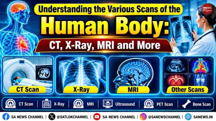
Do you ever realise that doctors can look into our body even without making an incision? Medical scans are the advanced technology that transforms the invisible structures and abnormalities into images that can guide diagnosis and treatment. From the most common X-ray to the advanced CT scans, PET scans and MRI, medical scans use a different method to reveal what is happening inside the human body.
- Common Types of Medical Scans
- CT vs X-Ray vs MRI vs Ultrasound: What Is the Difference?
- How Does Each Scan Function
- Image Quality and Degree of Detail
- Radiation Exposure
- Time Needed and Comfort of the Imaging Exam
- Which Scan Is Best for Different Parts of the Body?
- Are CT, X-Ray, MRI and Other Scans Safe?
- Which Scans Use Radiation?
- Safety of MRI and Possibility of Metal Implants
- Contrast Materials and Potential Hazards
- Taking into Account Risks and Benefits
- Medical Scans Can Detect Bodily Diseases, But One Disease Remains Beyond Their Reach
Medical scans are useful not only in the detection of fractures, tumors, infections and internal injuries of the body, but also to diagnose diseases and evaluate how well a treatment is working. Surprisingly, different imaging technology operates through different energy forms: MRI operates on radio and magnetic waves, while PET uses different tracers to detect the metabolic activity of the body.
Understanding how these scans work can help you know why a doctor may recommend one test over another.
Common Types of Medical Scans
Modern medicine adopts various medical imaging scans. Each imaging scan offers its own diagnostic features. Some show scans of the bones, while others provide intricate details of soft tissues or show various metabolic changes occurring within the body. Knowing the difference between various medical imaging scans makes it easier to understand why doctors recommend a particular imaging test.
X-Ray Scan
X-ray is one of the oldest and the most common medical imaging scans. It scans the dense structure of the body, such as bones by passing a small amount of ionizing radiation through the body. These scans are most commonly used to detect fractures, chest infections, dislocations, and certain lung conditions. They are generally quick and widely available, making them a common first-line imaging test.
CT Scan
CT (Computed Tomography) refers to the procedure which makes use of many X-ray pictures acquired from various directions which are processed through computer techniques to generate detailed images of the cross sections of the body. In contrast to the ordinary X-rays, CT scan gives a much more detailed view of the internal structures in the body such as organs, blood vessels, bones and also the soft tissues present inside. The process is even more useful for detecting the internal bleeding and injuries caused by trauma and also for identifying tumors and other defects.
MRI Scan
An MRI (Magnetic Resonance Imaging) adopts the use of non-ionizing strong magnetic and radio waves to create highly detailed images. MRI is specifically effective for examining soft tissues that may not be clearly visible on an X-ray. . One of the most important considerations is that MRI scanners can be noisy and may require patients to remain still inside a relatively enclosed space.
Ultrasound Scan
An ultrasound scan uses high-frequency sound waves to create real time images. Rather than X-rays, which use ionizing radiation, an ultrasound is widely used for examining the abdomen, pelvis, blood flow, soft tissues and developing fetus during pregnancy. Ultrasound also allows doctors to observe movement and blood flow while the examination is taking place. Its portability and lack of ionizing radiation make it a particularly versatile diagnostic imaging technique.
PET Scan and Bone Scan
PET (Positron Emission Tomography) scans and bone scans fall under the category of nuclear medicine imaging tools. These devices use small traces of radiopharmaceuticals to allow researchers to visualize and identify biological activities instead of structural information. They have unique capabilities to visualize how tissue works. PET scans, for example, can indicate areas where metabolism is high. PET scans are widely used in cancer detection, staging, and monitoring therapy success. Bone scans can indicate the areas of high or low bone activity.
CT vs X-Ray vs MRI vs Ultrasound: What Is the Difference?
The selection between a CT scan, X-ray, MRI or ultrasound depends on what doctors need to see inside the body. Although all four are medical imaging techniques, all of them use different technologies and provide different levels of imaging details. Understanding how each scan works, the type of images it produces, its use of radiation and the patient experience can explain why one test may be recommended over another.
How Does Each Scan Function
X-ray imaging employs ionizing radiation to generate images. A CT scan uses X-rays to capture images from various positions to form detailed cross-sectional images. An MRI relies on magnetic fields and radio waves while an ultrasound depends on sound waves to produce live images.
Image Quality and Degree of Detail
The level of detail varies considerably with the technique used to acquire an image. X-ray imaging may yield simple two-dimensional images that are helpful in imaging the chest and bones. CT imaging produces significantly more detailed cross-sectional images of the internal organs, bones, blood vessels and tissues. An MRI provides high-resolution soft tissue images, including those of the brain, spinal cord, muscles and ligaments. Ultrasound imaging is able to produce live images and is best suited to certain soft tissues, blood circulation and pregnancy diagnostics.
Radiation Exposure
Radiation exposure is one of the most significant differences among these scans. Both CT scans and X-rays utilize ionizing radiation; however, the amount of radiation is different depending on the type of the particular examination. CT scan usually has higher radiation exposure compared to a standard X-ray examination. MRI and ultrasound do not use ionizing radiation, which makes those imaging techniques fundamentally different. Nevertheless, any form of imaging has its advantages, disadvantages, and safety issues, so physicians select the method of imaging depending on a patient’s health condition.
Time Needed and Comfort of the Imaging Exam
The duration of the imaging examination is variable. X-ray examinations can be completed quickly, CT scan is fast too and is useful in emergency cases. An MRI scan is usually longer and requires a patient to lie still in the machine. The ultrasound imaging procedure is easy and pleasant.
Also read: COVID-19 के बाद नई बीमारियों पर रिसर्च: Long COVID, Disease X और भविष्य की महामारियों की तैयारी
Which Scan Is Best for Different Parts of the Body?
There is no single imaging test that is best for every body part. Doctors choose a scan based on the suspected condition, symptoms and type of tissue that needs to be examined.
Brain and Head
Doctors generally recommend CT scans during emergencies such as head injuries and suspected bleeding, while an MRI is generally suitable to get greater details of the brain and the soft tissues.
Bones and Joints
To detect fractures and other bone problems, X-rays are commonly used for medical imaging scans. CT provides more detailed views of complex bones, whereas MRI is specifically useful for cartilage, ligaments and other soft tissues around joints.
Chest and Lungs
For initial chest and lung evaluation, X-rays are commonly used. CT scans are used to get more detailed images of the lungs and surrounding structures.
Abdomen and Pelvis
Depending on the suspected condition, doctors may use ultrasound, CT or MRI to examine vital organs including the liver, kidneys, gallbladder and reproductive organs.
Muscles and Soft Tissues
MRI is mainly useful for detailed imaging of muscles, tendons, ligaments, nerves and other soft tissues.
Are CT, X-Ray, MRI and Other Scans Safe?
Medical imaging is generally considered safe when used appropriately. However, each technique has different benefits, limitations and safety considerations.
Which Scans Use Radiation?
While X-rays, CT scans and many nuclear medicine scans use ionizing radiation, ultrasound and MRI do not use ionizing radiation.
Safety of MRI and Possibility of Metal Implants
The MRI technology utilizes enormous magnetic forces for producing images. It is advisable for patients to notify the medical professionals about the presence of metal implants including pacemakers, clips, or other metallic objects which may create problems during the procedure.
Contrast Materials and Potential Hazards
Some tests like CT, MRI, and others rely upon the contrast substances to visualize certain body systems. The patients should always inform the medical experts about their history of allergies, kidney dysfunction, and their previous experiences with the contrast materials.
Taking into Account Risks and Benefits
Doctors should weigh the importance of the symptoms, medical history of the patient, and diagnostic value of proposed imaging before making a recommendation. The goal is to obtain useful information without any unnecessary procedures and risks.
Medical Scans Can Detect Bodily Diseases, But One Disease Remains Beyond Their Reach
While modern medical science uses diagnostic tools such as MRI, CT scans, X-rays, and PET scans to detect diseases and understand the physical condition of the human body, one disease still remains hidden.
In His spiritual teachings, Saint Rampal Ji Maharaj preaches that human life is precious, as every soul remains caught in the cycle of birth and death and experiences various forms of suffering.
Saint Rampal Ji Maharaj provides the knowledge that human life is the only form among the 84 lakh births, in which a soul seeks spiritual knowledge to attain liberation from the cycle of birth and death. Only Tatvadarshi Saint can provide true spiritual knowledge to get rid of the cycle of birth and death. To know about the Tatvadarshi Saint, visit www.jagatgururampalji.org or read the sacred book Gyan Ganga.